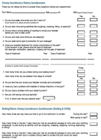
Office assessment of urinary incontinence By Lars Viktrup, MD, PhD, and Richard C. Bump, MD Urinary incontinence can often be successfully treated without referral, but first ob/gyns need to broach the subject! Two experts tell how to evaluate this widespread condition. Urinary incontinence (UI) is an underdiagnosed disorder that affects millions of women, causing substantial emotional and financial burdens to them and their families. Historically, women have been reluctant to report UI symptoms to their physicians; however, with increasing awareness of the disorder, attitudes are expected to change and more and more women may be consulting their ob/gyns for assessment and treatment. Our purpose is to review the major types of UI, techniques for assessing UI risk factors and patients' histories, recommended elements of a physical examination, and additional office-based tests of urinary function. We'll also address when referral for advanced diagnostic testing is appropriate. We suggest conducting the baseline assessment in every ob/gyn's office. It is fast, safe, inexpensive, and sufficient for initiating treatment in most women with UI. UI is prevalent, bothersome, and expensive. In a North American survey of 24,581 noninstitutionalized women, 37% reported UI during the previous month and almost half of those with incontinence said the symptom was moderately to extremely bothersome. 1 Similarly, a recent international review found that UI affects more than 30% of all women, 2 a rate that rivals that of chronic diseases like hypertension or diabetes. 3-5 The National Institutes of Health has estimated the total annual direct cost of UI (encompassing everything from the cost of disposable products to long-term-care facilities) as higher than the total annual cost for diseases such as osteoporosis and arthritis. 6 Even though the condition bothers them, only half of women with UI discuss it with their physician, while the remainder seem to accept UI as a natural consequence of growing older. 1 This may be partly due to the high UI incidence after childbirth. 7 In fact, some women with mild or fluctuating UI may experience little impact on their quality of life, with no need for treatment and may be satisfied with managing the condition themselves with absorptive products such as pads. In contrast, others may be too embarrassed to talk about UI, believe nothing can be done, or think an undesired surgical procedure is their only treatment option. 1,8,9 Unfortunately, years may elapse between the onset of UI symptoms and a woman's seeking treatment, usually with a family practitioner, a gynecologist, or an internist. 1 More women might discuss their condition earlier if physicians routinely raised the subject of UI during exams, but few physicians do. 10 After a simple assessment, UI can often be treated successfully in the ob/gyn office; however, it's proven difficult to change existing habits. 11-13 A survey of noninstitutionalized women with UI found that limiting fluid intake was the most frequent coping technique (38%). 1 The Agency for Healthcare Research and Quality (AHRQ) and the International Consultation on Incontinence (ICI) recommend that a basic assessment of women with UI include: the history of its severity and impact on quality of life, a gynecologic/neurologic examination, a urinalysis, a frequency/volume diary, a stress- or cough pad-test, and, if needed, assessment of post-void residual urine (PVR) volume. 14-16 The entire algorithm has not been evaluated for use in all women with different types of UI, but single elements have been tested and validated. 16-18 For most patients with UI who see their ob/gyn for initial care, this assessment would suffice to start low-risk, reversible, and inexpensive lifestyle changes, behavioral modification, or approved pharmaceutical treatment. Even though some women may not be ideal candidates for the treatment selected, the most efficient use of resources might be a nonsurgical trial, followed by an assessment of their responses. Since any UI assessment should be tailored to individual patients' symptoms and individual physicians' expertise, this article evaluates the options for initial UI assessments that can be conducted in an ob/gyn setting, focusing on the AHCPR's recommendations in 1996 and ICI 2002. A literature search for scientific evidence supporting the recommendation was performed using PubMed, the Cochrane Database, the AHCPR (former name of the AHRQ) 1996 recommendations, and ICI 2002 Incontinence Book. Some of the suggested questionnaires presented here (S/UIQ, SUBS scale) are recently developed and are being validated in ongoing studies. Urinary incontinence, which is the accidental leakage of urine onto clothing, underwear, or pad, differs in type and severity (see "Distinguishing the types of urinary incontinence" and Table A). Obtain a detailed medical history An important part of evaluating and classifying UI, the detailed medical history should focus on types of incontinence symptoms, patterns of voiding, quality of life, co-existing medical conditions, and precipitating factors. 19 Also consider (when appropriate) the patient's mental status, mobility, and living environment because urine loss may stem from factors other than malfunction of the lower urinary tract. But regardless of the care taken in obtaining a UI history, patient-provided information about the onset of UI may be unreliable. 7 With that in mind, special emphasis should be directed to potentially reversible factors, especially in elderly patients. The acronym DIAPPERS (Delirium/confusional state, Infections, Atrophic urethritis/vaginitis, Pharmaceuticals, Psychological, Excessive urine output, Restricted mobility, Stool impaction) has been suggested as a way to remember these transient factors, but delirium is rare and you should also consider other potential risk factors such as obesity, coughing, and pelvic organ prolapse (POP). 20 To standardize the collection of information, certain instruments may be used as described below. Additionally, the patient and the nursing staff may complete part of it before the first consultation with the physician. UI history questionnaire. Before the consultation, the patient can complete a standardized questionnaire about her symptoms, previous surgeries, medications, and related dates. Such a questionnaire can elicit valuable information the woman may have otherwise forgotten, and save considerable time during the office consultation. The UI history questionnaire (the first questionnaire shown) is one that has been used successfully by women with UI before a scheduled urogynecology consultation at Duke University . However, an ob/gyn's personal preference for certain information should be considered.
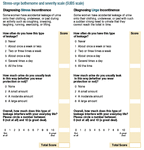
Voiding and UI symptom assessment questionnaire. A patient can also complete a questionnaire about voiding habits and leakage prior to a consultation, and before and/or after treatment (the second questionnaire). The stress urge incontinence questionnaire (S/UIQ) currently being validated within clinical trials conducted by Lilly may prove to be an easy way to discriminate between SUI and UUI. It is considered normal to void seven times daily and up to two times during the night. Frequency is defined as the individual complaint of too many voids during waking hours, while nocturia is the complaint of waking at night one or more times to void. 16
Quality-of-life questionnaire. Since UI can significantly impact quality-of-life (QOL) a QOL questionnaire can help monitor the initial severity and treatment response. 21 A good QOL score is not just the absence of a disease, but relates to the multidimensional approach to evaluating a given condition's impact on general health. 22 It evaluates such key areas as psychosocial impact, avoidance and limiting behavior, and social embarrassment. 23 Of course, it's not possible for a short questionnaire appropriate for use in a busy ob/gyn office to fully address all these areas. Therefore, some experts suggest shorter condition-specific scales for rating bothersomeness. The International Consultation on Incontinence has suggested using the Quality-of-Life Short Form questionnaire, which measures the severity of UI and its interference with everyday life. 19 This instrument's reliability is excellent and its correlation with the ICI pad test is reasonable, but it is still undergoing validation. 19 It does not discriminate between the bothersomeness caused by different types of UI. Since it has been recommended to treat the more bothersome symptom first in patients with mixed urinary incontinence, this questionnaire's lack of discrimination may be an important limitation. 24 Therefore, researchers are currently testing and validating a similar instrument that separates the bothersomeness of SUI from that of urge urinary incontinence. Real-time symptom diary A more accurate and detailed picture of a patient's symptoms can be obtained using a real-time symptom diary. Figure 1 gives an example of a 7-day voiding diary, but a 4-day diary is equally reliable. 25,26 It is recommended that this diary be supplemented with information about the time, type, and amount of daily fluid intake (especially caffeine and alcohol consumption), as well as related events or physical activity during the course of the day. 16 A patient-friendly symptom diary incorporating these features can be downloaded at http://kidney.niddk.nih.gov/kudiseases/pubs/diary/ , the Web site of the National Institute of Diabetes & Digestive & Kidney Diseases (NIDDK). 27 A similar diary can also be seen on the American Family Physician Web site ( http://www.aafp.org/afp/20001201/2433.html ). 27
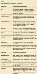
PLEASE NOTE: If you are using Internet Explorer version 6 or higher, after clicking to enlarge the graphic, click the picture icon that appears in the lower right-hand corner of the graphic. SUI history, bladder neck hypermobility, or intrinsic sphincter deficit. A typical history of simple SUI not associated with severe urethral sphincter failure (intrinsic sphincteric deficiency or ISD) would include a single-spurt loss of urine coinciding with a physical activity that causes an abrupt rise in abdominal pressure (like a cough or sneeze), and without predominant UUI, urgency, frequency, bedwetting, or nighttime frequency. More prolonged loss that starts seconds after the physical stress suggests stress-induced detrusor overactivity. Patients who are more likely to have SUI with ISD are those who note continuous UI with negligible stress in any position, while those more likely to have UUI are those who have nocturnal enuresis or advanced-stage pelvic organ prolapse visible beyond the vaginal opening. Other patients with less chance of simple SUI are African-American women, nulliparous women, and women with previous failed continence surgery. 29 History and surgical failure. After continence surgery, patients with a history of urinary retention or frail elderly patients have an increased risk of complications, such as retention and overflow UI. Women with increased risk of SUI reoccurring after continence surgery include patients with ongoing excessive stresses to the pelvic floor (for example, constipation, obesity, unusual occupational or recreational stress, smoking, and chronic obstructive pulmonary disease (COPD)) and women with severe pelvic floor neuropathy or myopathy. 30,31 Risk factors A number of risk factors have been associated with both female SUI and UUI (Table 1). 2,32-43 These risk factors from the patient's history and objective findings should be assessed during the UI consultation and efforts should be directed toward eliminating the transient factors and avoiding the ongoing risk factors. The main inciting factor, though, is often unknown, since UI may have a fluctuating nature and multiple causes.
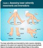
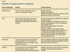
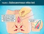
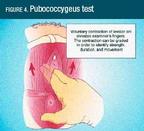
Physical laboratory examination A focused physical examination of a woman with UI includes abdominal/rectal, pelvic, and neurologic examinations. Figure 2 shows how to test lower extremity movements and innervation (lumbar-sacral cord), while Figure 3 illustrates the bulbocavernosus reflex test (sacral cord). Interpret these tests carefully, however, because an absent sacral reflex does not definitely diagnose a neurogenic lesion, and its presence does not exclude a partial lesion. 44 Urinalysis is also recommended to rule out local lower urinary tract disorders, especially infection (Table 2). The strength of the pelvic floor's pubococcygeus muscle may be assessed using a 9-point scale based on the strength, duration, and perineal movement observed with a muscle contraction (Figure 4). Table 3 is adapted from one initially described by Brink and colleagues . 45 Although the method is subjective and not sensitive, it's useful to determine treatment effect in the same patient. 46 A similar muscle grading may be applied to the anal sphincter.
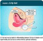
Office-based or -initiated tests of urinary function Because urodynamic equipment is not usually available in the ob/gyn's office, and since the results of urodynamic studies do not always explain symptoms, 47-49 UI is often initially managed without urodynamics, which should be reserved for complicated cases, after treatment failure, or prior to invasive, irreversible, or highly specific treatment. 50,51 Controversy exists about the use of urodynamics in patients with symptoms of overactive bladder (OAB); some suggest urodynamics in all patients with OAB symptoms, while others reserve such studies for patients with persistent symptoms, 52,53 or before considering continence surgery. 18 While the stress pad test, cough stress test, and PVR urine measurements are recommended for the basic evaluation of all women with UI, the Q-tip test and eyeball cystometry may be reserved for special cases (Figures 5 and 6).
Pad test. A short-term pad test conducted in the office, or a long-term version performed at home are used for quantitative measurements of UI, although controversy exists about their reproducibility and validity. 17,54 The short-term pad tests should include standard procedures, preferably at a fixed bladder volume (for example, bladder filling by catheterization and/or confirmed with ultrasound). 17 Ideally, this test should be done when a patient's bladder is full but before she has a strong urge to void. 55 The test measures the amount of leakage that occurs as the patient performs a series of activities that might trigger urine leakage. There are a variety of recommended activities (for example, walking for 30 minutes; including climbing up and down the equivalent of a flight of stairs), going from standing to sitting position 10 times, coughing vigorously 10 times, running in place for 1 minute, bending to pick up a small object from the floor five times, and washing one's hands in running water for 1 minute. These activities are more indicative of SUI, except for leakage of urine when washing hands in running water for 1 minute, which is more suggestive of UUI. A gain in pad weight of 1 g or more is considered to be a positive 1-hour test. The sequence of activities has little effect on test results. 54 A 24- to 48-hour pad test performed in familiar surroundings may have better reproducibility, but makes the stress component of the leakage less certain. 17 The woman is supplied with pre-weighted pads and a plastic bag in which to store the used ones. A pad weight gain of 4 g or more is considered to be a positive 24-hour test. 55 Cough stress test. You can document SUI by having the patient cough forcefully while in a supine, sitting, or standing position at the end of bladder filling. After removal of the catheter, the ob/gyn observes leakage from the urethra at the instant she coughs. Post-void residual urine volume . PVR is considered significant if the amount of urine remaining in the bladder after voiding exceeds 30% of the total bladder capacity (usually >50 to 100 mL), and may indicate bladder outlet obstruction, detrusor underactivity, or both. 15 PVR should be assessed using catheterization or ultrasound, because abdominal palpation is unreliable. 56 Q-tip test. A test with a cotton-tipped swab (Figure 5) is a simple inexpensive way to identify the bladder neck hypermobility often associated with SUI. Insert a sterile, lubricated, and solid cotton tip swab into the urethra until it lies just within the urethrovesical junction, usually 1 inch from the urethral opening. Using a goniometer, measure the maximum angle deflection from horizontal while the woman is performing a maximum Valsalva effort. Urethrovesical hypermobility is typically diagnosed when the bladder neck moves downward, posteriorly causing the straining Q-tip angle to exceed 30° above the horizontal. 57 Eyeball cystometry. Simple bladder filling or "eyeball cystometry" (Figure 6) is retrograde filling of the bladder through a catheter in an effort to identify abnormal bladder activity or sensation. Instilling sterile water or saline through a catheter into the bladder will normally induce a first sensation of the need to void after 100 to 200 mL and indicate a maximal bladder capacity of 350 to 600 mL. Severe urgency occurring below a 300 to 400 mL bladder volume suggests sensory urgency, whereas unsuppressed bladder contractions (identified by a raise in the fluid level in the funnel) suggests detrusor overactivity. This detection of detrusor overactivity is not substantially different in simple versus electronic cystometry, 51,58-61 however, abdominal straining as a cause of the pressure rise can be difficult to exclude without multichannel electronic studies. Indications for further evaluation Most often, UI can be categorized and presumptively treated by the ob/gyn based on a patient's history of symptoms and a selection of the assessments previously described. For some patients, however, referral for further evaluation may be appropriate. The ICI 50 and AHCPR 15 recommend advanced diagnostic testing if the diagnosis is uncertain (for example, lack of correlation between symptoms and clinical findings) or if it is not possible to develop a reasonable treatment plan using the basic diagnostics. Additional situations where referral may be appropriate are: • failure to respond—to the patient's satisfaction—to an adequate therapeutic trial, and the patient is interested in pursuing further therapy; • consideration of surgical intervention, particularly if a previous surgery failed or the patient is a high surgical risk; • suspected urinary fistula; • hematuria or pain without infection; • the presence of comorbid conditions, such as: • incontinence associated with recurrent symptomatic UTI, • persistent symptoms of difficult bladder emptying, • history of previous continence surgery, radical pelvic surgery, or pelvic irradiation, • symptomatic externally visible POP, • abnormal PVR, • related neurologic conditions, • suspected malignancy. Local referral patterns and requirements may also dictate the circumstances determining further evaluation and treatment. Advanced diagnostic testing can include uroflowmetry, cystourethroscopy, multichannel urodynamics, videourodynamics, electrophysiologic testing, urethral pressure profilometry, and Valsalva leak point pressure testing. 51 Conclusions Although ob/gyns may see the initial assessment of women with UI as involved and time consuming, this article identifies different ways of reducing the complexity of evaluation. Ongoing testing and validation of simple questionnaires presented here may result in simpler tools that are sufficient to initially assess female UI before low-risk treatment trials.
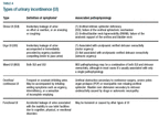
Distinguishing the types of urinary incontinence UI is described at four levels: (1) the symptom is a subjective description of the condition by the patient; (2) the sign is the documentation by the physician; (3) it is the finding during urodynamic studies; and (4) it is the composite diagnosis based on urodynamic observations associated with characteristic symptoms and signs of UI. 1 From a recent study of noninstitutionalized North American women with UI, 41% reported only stress urinary incontinence (pure SUI) symptoms, 11% only urge urinary incontinence (pure UUI) symptoms, 45% mixed stress and urge incontinence symptoms (MUI), and 3% "other" incontinence. 2 This distribution is similar to that found in epidemiologic studies. 3 However, while history can elucidate the type and severity of UI, symptoms can't be used to make a definitive diagnosis of the underlying condition. When the UI symptoms of noninstitutionalized Norwegian women were re-evaluated by a gynecologist assessing symptoms, clinical evaluation, and urodynamics, the prevalence of SUI increased from 51% to 77%, MUI fell from 39% to 11%, and pure UUI remained stable at 10% to 12%. 4 Similarly, overactive bladder (OAB) symptoms like UUI, urgency, and frequency were poor predictors of detrusor overactivity when urodynamics were performed in 843 consecutive women at Kings College Hospital in London, UK. 5 In a retrospective study of 950 women with UI referred for urodynamics at Duke University (North Carolina), 52% of the patients evaluated had symptoms of MUI, but only 14% had the conditions of both urodynamic stress incontinence (USI) and detrusor overactivity when tested. 6 The majority went from mixed symptoms to pure USI. Among the women with pure SUI symptoms, however, 11% did not have the SUI confirmed urodynamically. 6 PLEASE NOTE: If you are using Internet Explorer version 6 or higher, after clicking to enlarge each graphic in the article, click the picture icon that appears in the lower right-hand corner of the graphic.
|
||||
